Boil
Importance of Early Diagnosis of HS
Many patients experience significant delays in receiving a diagnosis of hidradenitis suppurativa (HS). This is often due to1,2:
- Patients waiting to seek medical attention until later stages of disease
- HS being mistaken for other conditions with similar symptoms
Prompt, accurate diagnosis can aid in the initiation of proper treatment and help prevent the progression to more debilitating stages.1,3
HS Diagnosis Statistics
Patients experience a significant delay in receiving an HS diagnosis
Average time to diagnosis is 7-10 years4
Patients are initially misdiagnosed
Patients may have more than 17 visits before receiving a correct diagnosis5
Patients may visit multiple clinicians before receiving a diagnosis
4 to 5 physicians may be seen prior to an accurate HS diagnosis4-6
Patients may initially think symptoms are temporary and not too bothersome
Patients wait an average of 2.3 years before first presenting to a physician6
Differential Diagnoses of HS
Follicular pyodermas (carbuncles, furuncles, boils)7-9
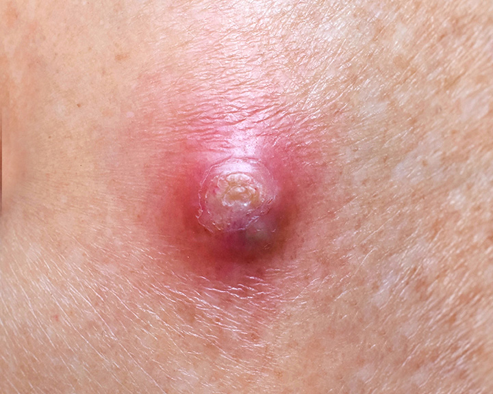
Differentiation: Unlike HS, which typically exhibits a chronic and recurrent course, follicular pyodermas are transient lesions that usually respond rapidly to appropriate antibiotic therapy. In addition, the follicular pyodermas do not cause the comedones, persistent sinus tracts, and hypertrophic scarring observed in HS.
Cutaneous Crohn’s disease7,10
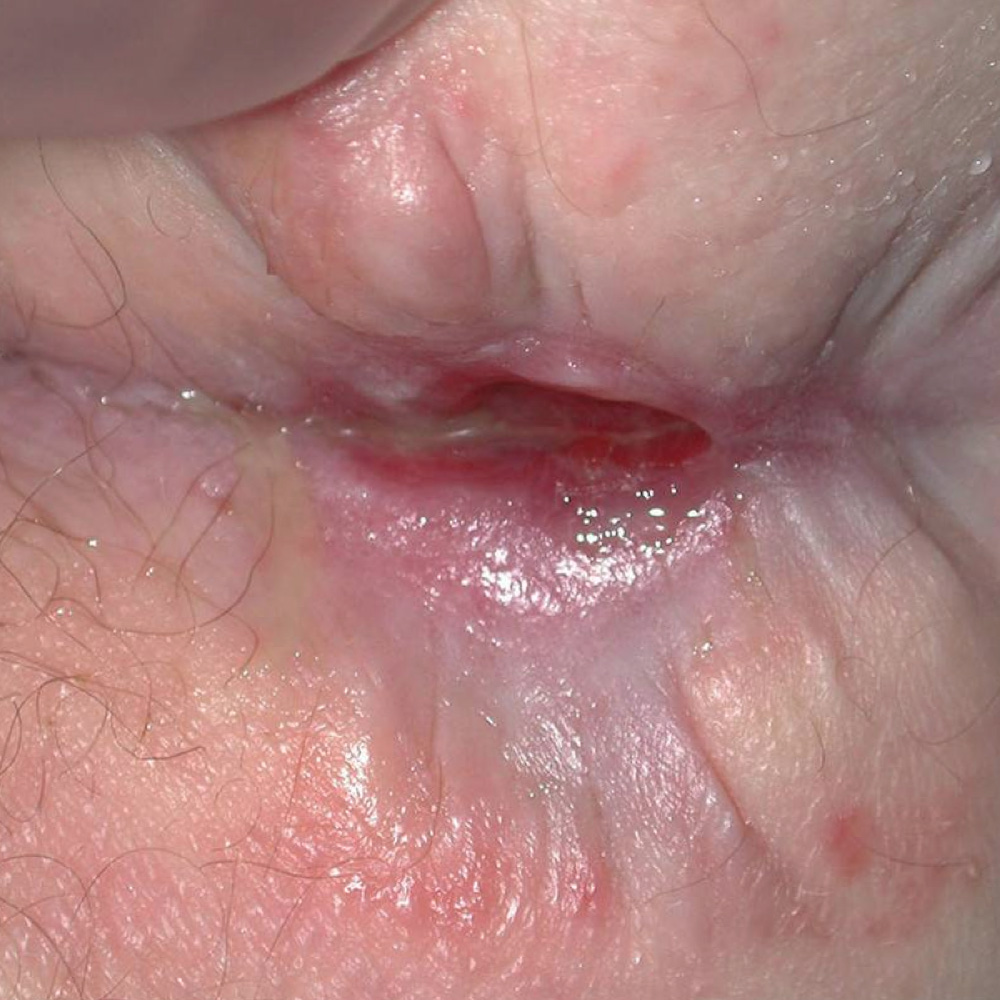
Cutaneous Crohn's disease
Adapted with permission from Rice SA, Woo PN, El-Omar E, et al. BMC Res Notes. 2013.10

This work is licensed under a Creative Commons Attribution 4.0 International License.
Differentiation: “Knife-cut” ulcers and no blackheads; fistulas connect with the gastrointestinal tract; concurrent with gastrointestinal Crohn’s disease.
Acne, cystic acne7
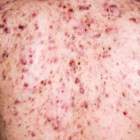
Cystic acne
Differentiation: Distribution on the face, back, and upper chest, and include whiteheads. HS primarily involves the axillae, groin, buttocks, and inframammary folds. Additionally, HS typically involves more deep-seated lesions leading to sinus tracts and scarring.
Granuloma inguinale7
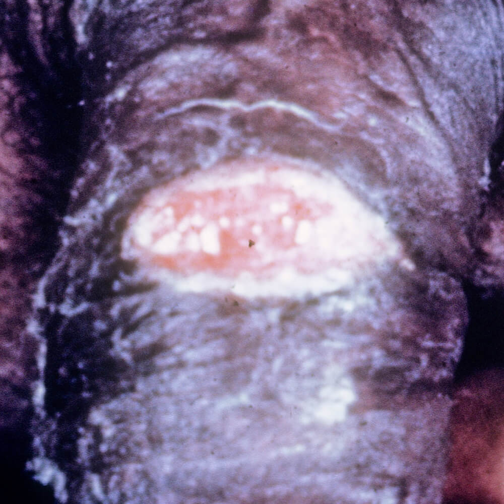
Granuloma inguinale
Differentiation: Red ulcers that bleed easily, granulation of tissue; has Donovan bodies (histology); infectious agent: Klebsiella granulomatis.
Lymphogranuloma venereum7
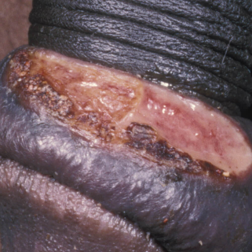
Lymphogranuloma venereum
Differentiation: Bacterial etiology: Chlamydia trachomatis (serotype L1-L3)
Actinomycosis7
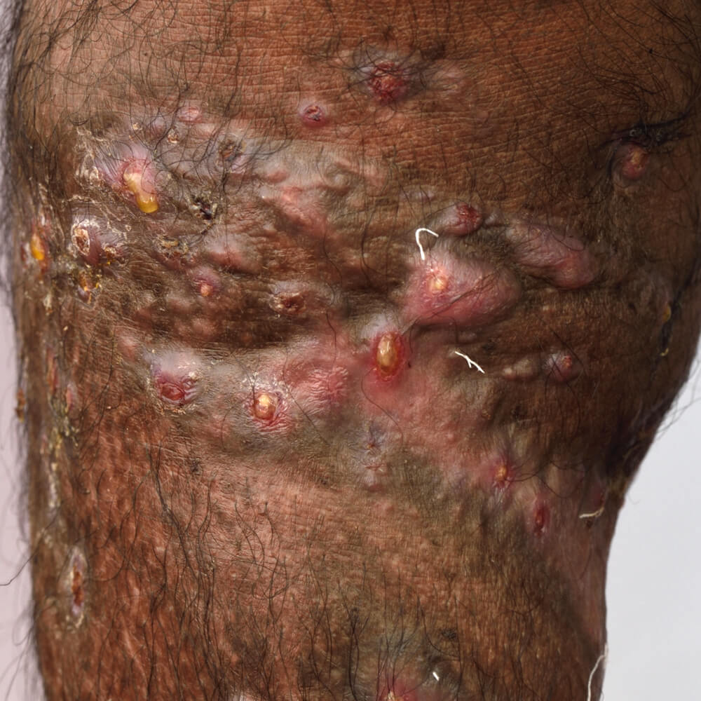
Actinomycosis
Differentiation: Bacterial infection caused by Actinomyces
Cutaneous tuberculosis7
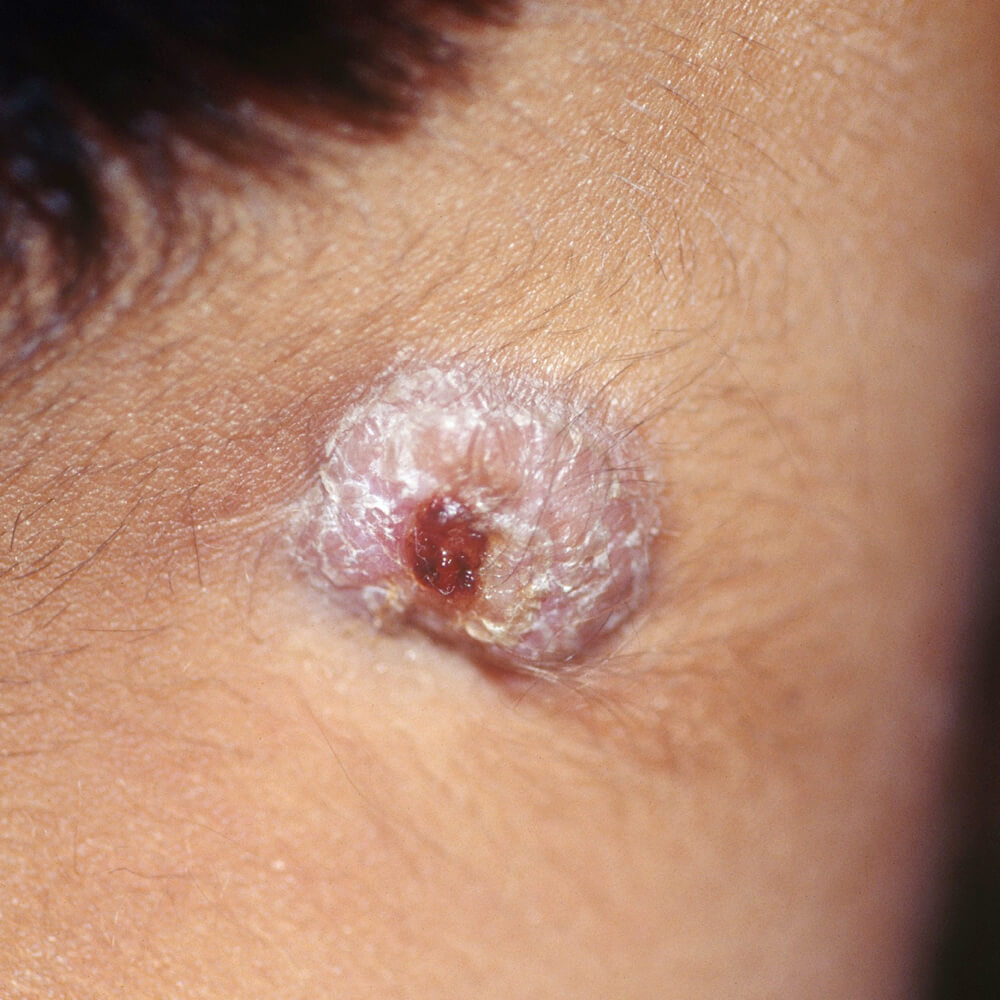
Tuberculosis verrucosa cutis
Differentiation: Bacterial infection caused by Mycobacterium
Other considerations11
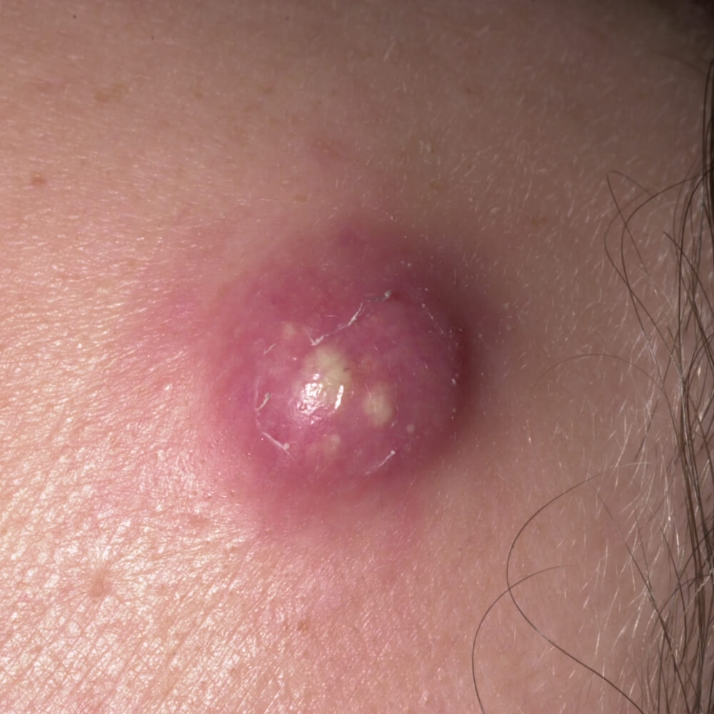
Epidermoid cyst
- Epidermoid or dermoid cyst12
- Pilonidal cyst
- Erysipelas
Review the following 3 patient cases and determine the most likely diagnosis. Reference the HS diagnosis criteria to help inform your choices.
Choose your specialty:
“It’s critically important to diagnose HS early and intervene with medical therapies given the progressive, irreversible nature of the disease.”
- Dermatologist
REFERENCES
1. Micheletti RG. Natural history, presentation, and diagnosis of hidradenitis suppurativa. Semin Cutan Med Surg. 2014;33(suppl 3):S51-S53. 2. Jemec GBE. Hidradenitis suppurativa. N Engl J Med. 2012;366(2):158-164. 3. Kimball AB, Rahawi K, Duan Y, Alavi A, Okun MM. Impact of delayed diagnosis in patients with hidradenitis suppurativa (HS): real-world data from the UNITE HS registry. Poster presented at: Symposium on Hidradenitis Suppurativa Advances; November 1-3, 2019; Detroit, MI. 4. Garg A, Neuren E, Cha D, et al. Evaluating patients' unmet needs in hidradenitis suppurativa: results from the Global Survey Of Impact and Healthcare Needs (VOICE) Project. J Am Acad Dermatol. 2020;82(2):366-376. 5. Alavi A, Lynde C, Alhusayen R, et al. Approach to the management of patients with hidradenitis suppurativa: a consensus document. J Cutan Med Surg. 2017;21(6):513-524. 6. Saunte DM, Boer J, Stratigos A, et al. Diagnostic delay in hidradenitis suppurativa is a global problem. Br J Dermatol. 2015;173(6):1546-1549. 7. Saunte DML, Jemec GBE. Hidradenitis suppurativa: advances in diagnosis and treatment. JAMA. 2017;318(20):2019-2032. 8. Boils and carbuncles. Mayo Clinic. Updated September 11, 2019. Accessed January 29, 2020. https://www.mayoclinic.org/diseases-conditions/boils-and-carbuncles/symptoms-causes/syc-20353770. 9. Collier F, Smith RC, Morton CA. Diagnosis and management of hidradenitis suppurativa. BMJ. 2013;346:f2121. 10. Rice SA, Woo PN, El-Omar E, Keenan RA, Ormerod AD. Topical tacrolimus 0.1% ointment for treatment of cutaneous Crohn's disease. BMC Res Notes. 2013;6:19. 11. Shah N. Hidradenitis suppurativa: a treatment challenge. Am Fam Physician. 2005;72(8):1547-1552. 12. Dermnet Skin Disease Atlas. Accessed June 18, 2019. http://www.dermnet.com/images/Epidermal-Cyst/picture/16454.
